Using tiny semiconductor crystals, biological probes
and a laser, Johns Hopkins engineers have developed a new
method of finding specific sequences of DNA by making them
light up beneath a microscope. The researchers, who say the
technique will have important uses in medical research,
demonstrated its potential in their lab by detecting a
sample of DNA containing a mutation linked to ovarian
cancer.
The Johns Hopkins team described the new DNA
nanosensor in a paper published in the November issue of
the journal Nature Materials.
"Conventional methods of finding and identifying
samples of DNA are cumbersome and time-consuming," said
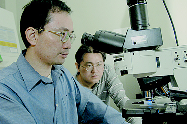
Jeff Tza-Huei
Wang, senior author of the paper and supervisor of the
research team. "This new technique is ultrasensitive, quick
and relatively simple. It can be used to look for a
particular part of a DNA sequence, as well as for genetic
defects and mutations."
The technique involves an unusual blend of organic and
inorganic components. "We are the first to demonstrate the
use of quantum dots as a DNA sensor," Wang said.
Quantum dots are crystals of semiconductor material
whose sizes are only in the range of a few nanometers
across. (A nanometer is one-billionth of a meter.) They are
traditionally used in electronic circuitry. In recent
years, however, scientists have begun to explore their use
in biological projects.
Wang, an assistant professor in the
Department of Mechanical
Engineering and the
Whitaker Biomedical
Engineering Institute at Johns Hopkins, led his team in
exploiting an important property of quantum dots: They can
easily transfer energy. When a laser shines on a quantum
dot, it can pass the energy on to a nearby molecule, which
in turn emits a fluorescent glow that is visible under a
microscope.
But quantum dots alone cannot find and identify DNA
strands. For that, the Johns Hopkins team used two
biological probes made of synthetic DNA. Each of these
probes is a complement to the DNA sequence the researchers
are searching for. Therefore, the probes seek out and bind
to the target DNA.
Each DNA probe also has an important partner. Attached
to one is a Cy5 molecule that glows when it receives
energy. Attached to the second probe is a molecule called
biotin. Biotin sticks to yet another molecule called
streptavidin, which coats the surface of the quantum
dot.
To create their nanosensor, the researchers mixed the
two DNA probes, plus a quantum dot, in a lab dish
containing the DNA they were trying to detect. Then nature
took its course. First, the two DNA probes linked up to the
target DNA strand, holding it in a sandwichlike embrace.
Then the biotin on one of the probes caused the DNA
"sandwich" to stick to the surface of the quantum dot.
Finally, when the researchers shined a laser on the
mix, the quantum dot passed the energy on to the Cy5
molecule that was attached to the second probe. The Cy5
released this energy as a fluorescent glow. If the target
DNA had not been present in the solution, the four
components would not have joined together, and the
distinctive glow would not have appeared. Each quantum dot
can connect to up to about 60 DNA sequences, making the
combined glow even brighter and easier to see.
To test the new technique, Wang's team obtained DNA
samples from patients with ovarian cancer and detected DNA
sequences containing a critical mutation. "This method may
help us identify people at risk of developing cancer, so
that treatment can begin at a very early stage," Wang
said.
Lead author on the Nature Materials paper was
Chun-Yang Zhang, who was a postdoctoral fellow in Wang's
lab when the research was conducted. Co-authors were
Hsin-Chih Yeh, a doctoral student in the Department of
Mechanical Engineering, and Marcos T. Kuroki, who was an
undergraduate majoring in
biomedical
engineering when the research was conducted.
Funding for the research was provided by the National
Science and Whitaker foundations.
The university has filed for a provisional patent
covering the DNA nanosensor technology.


 GO TO
GO TO