 |
|


| Overview | Engineered nanoparticles for gene delivery | Biomimetic nanofibers for stem cell culture and tissue regeneration |
|
Engineered nanoparticles for gene delivery The development of safe and efficient carriers for in vivo gene transfer has been one of the key challenges in fulfilling the promises of gene therapy. DNA-compacting nanoparticles have been developed as a safer alternative to viral vectors, due to the ease of synthesis of these polycation/DNA nanoparticles, flexibility in the size of the transgene to be delivered, and minimal host immune responses induced by the carriers. However, these nanoparticles face serious challenges for in vivo applications, particularly when administered though intravascular administrations. Currently-available polymer/DNA nanoparticles, although showing excellent transfection efficiencies in vitro, often have negligible transfection activities in target tissue or cells of interest. This is mostly due to the severe aggregation of the complexes (poor colloidal stability) and in some cases dissociation of complexes in physiological media with high concentrations of serum and salts (poor complex stability). Specifically for liver-targeted delivery, the aggregated nanoparticles prevent the passage through sinusoid fenestrae and access to parenchymal cells, and lead to sequestration by the reticuloendothelial system. Therefore, developing nanoparticles with uniform sizes, high colloidal stability and high complex stability is the critical first step in developing an efficient liver-targeting strategy. We are working on several strategies towards this goal. New polymer carrier design that allows for controlled self-assembly of DNA/RNA nanoparticles
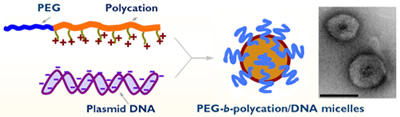
Figure 1. Illustration of micelle formation from PEG-b-polycation and DNA. The insert shows a TEM image of two PEG12K-b-PPA510K/DNA micelles (bar = 100 nm). We have developed a series of biocompatible and biodegradable polymers that self-assemble with DNA or RNA to form micellar nanoparticles. These nanoparticles consist of a DNA/polyphosphoramidate complex core and a polyethylene glycol (PEG) corona rendering particles more stable in aqueous medium. These nanoparticles are formed by self assembly, thus have uniform size ranging from tens to a couple hundreds of nanometers. With small and uniform size, good colloidal stability and longer circulation time, these nanoparticles offer opportunity for passive targeting to fenestrated tissues, for example solid tumor and liver sinusoid due to the “enhanced permeability and retention” effect. The size, morphology and stability can be controlled by adjusting PEG-b-PPA polymer structure. In addition, cell-specific ligands can be introduced to micelle surface to improve cell binding and targeting. Our micelle system offers unique advantages on versatile structure, biophysical properties, transfection efficiency and safety. These favorable characteristics make this micelle system an ideal platform for systematic investigation of delivery mechanism and optimization of tissue-targeted delivery system. An ability to control nanoparticle stability, DNA or RNA release, cell binding, and intracellular trafficking will be the key to understanding the rate-limiting steps for gene delivery and to optimizing transgene expression efficiency in vivo. We are currently investigating the effect of PEG-b-PPA carrier structure on DNA compaction ability, nanoparticle assembly and stability, transport properties at cell and tissue levels, and delivery efficiency. References:
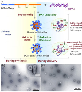
Dual-sensitive micellar nanoparticles to regulate DNA unpacking
References:
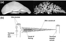
Figure 3. Human biliary system. (a). Voxel gradient display of the liver surface (left) and a computer-generated cholangiographic view of a normal human biliary tree (right), based on contiguous 2-mm slices imaged with electron-beam computed tomography. (b). Schematic representation of the terminal portions of the biliary tree showing a terminal bile duct with several draining bile ductules. The intralobular biliary system consists of bile canaliculi, which are 0.5- to 1.25-μm-wide spaces formed between adjacent hepatocytes. Liver-targeting strategies for nanoparticle delivery The liver is an important target for gene medicine applications. Successful liver-targeted gene delivery methods will be critical for treating liver-associated metabolic genetic disorders, viral infection and malignancies, and will also enable the expression of therapeutic products in the liver for treating systemic or off-site diseases. Intravenous and intraportal injections have been the primary routes for delivering nanoparticles or complexes to the liver. Even though intraportal injection provides a more direct delivery of nanoparticles to the liver, both methods face serious challenges in terms of particles being scavenged by Kupffer cells lining the sinusoidal endothelium and poor access to liver parenchymal cells. Retrograde intrabiliary infusion (RII), on the other hand, offers several unique advantages including direct access to hepatocytes and limited exposure to Kupffer cells. The biliary system consists of 7 to 10 orders of cholangiographically visible bile ducts spanning the expanse of the hepatic parenchyma (Fig. 3) with a surprisingly large and distensible volume (~29 ml for human liver). The ultrastructural surface of an entire normal biliary tree for a human liver would be around 3,000 cm2. This large surface area and broad distribution of the biliary system provide great access to nearly all hepatocytes in liver parenchyma through bile canaliculi, before the delivered drug or gene is exposed to KCs. Due to these advantages, we have been evaluating the feasibility of using RII as an effective administration route for liver-targeted gene delivery of nanoparticles. We have demonstrated that RII delivery strategy can overcome several key limitations of intravenous delivery and yield significantly higher levels of transgene expression in the liver. We have been focusing on optimization of administration parameters for RII of DNA nanoparticles, characterization of nanoparticle transport kinetics following RII as compared with intravenous injection, and understanding the mechanism by which DNA nanoparticles transfect liver parenchymal cells and other cells and tissue. The uniform size and high stability of PEG-b-PPA/DNA micellar nanoparticles in bile-containing medium make them excellent candidate for delivering genes through RII. We have been evaluating the efficiency and mechanism of gene delivery to rat and mouse livers using these micellar nanoparticles. Micelle formulations and delivery parameters will be optimized to achieve high and persistent gene expression. This will demonstrate the broad utility of this delivery strategy for expression of proteins intended for systemic distribution and for localized liver-specific diseases. References:
|