
Endbulb Research in the News
Scripps Howard News Service - December 1, 2005
ScienceNOW Daily News - December 2, 2005
New York Times News Service - December 2, 2005
London Telegraph - December 29, 2005
Endbulbs, Activity, and Cochlear Implants
Our ability to discriminate between various sounds requires the brain to be capable of identifying and separating the physical features of sound and then organizing them into different groupings to form a conscious precept. The physical attributes of sound include frequency composition, onset, duration, the time between sounds, and loudness. The precept could be the conversion of a string of utterances into understandable speech. Our ability to perform this function means that the brain is arranging different sounds (e.g., vowels and consonants) into a coherent pattern. How does it perform this task?
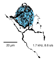
We have shown that endbulb morphology (arborization complexity, Sento and Ryugo, 1989) and synapse structure (synaptic vesicle number, mitochondrial volume fraction, postsynaptic density size, Ryugo et al., 1996) is systematically related to average levels of spike discharges. The implication is that in deafness where there is no spike activity, there should even more exaggerated synaptic change. How does deafness affect the development of structure and function of these terminals?
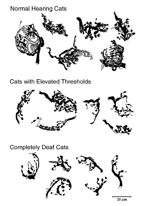
HRP-labeled endbulbs from the anteroventral cochlear nucleus.
Note the decreased branching in endbulbs of deaf and hard-of-hearing
cats. These abnormalities appear at about 6 months of age.
For congenitally deaf humans, the cochlear implant (or artificial ear) has been a very effective treatment. In many instances, children receiving a cochlear implant who also get prolonged audiologic training, gain functional "hearing" and speech. The question, however, is why are there some patients who fail to gain benefit from these devices? Does the abnormal appearance of endbulbs in deaf animals predict trouble?
Congenital deafness has been shown to cause neuronal death, induce abnormal circuits, and create synaptic abnormalities. Endbulbs of Held have been described in both deaf and hearing cats. In the electron microscope, it appears as a large profile adjacent to the cell body of a spherical bushy cell. In hearing cats, the profile is filled with large round synaptic vesicles. The apposed membrane is often interrupted by intercellular cisternae. The membrane apposition can be marked by a thickening on the bushy cell side and a concavity facing the endbulb. This thickening houses the transmitter receptors and is called the postsynaptic density (PSD). Endbulbs in deaf animals exhibited fewer synaptic vesicles, an absence of intercellular cisternae, and PSDs were larger and lost their curved appearance. Such pathologic changes may underlie the failure of some cochlear implant recipients to benefit from the devices. Children who are implanted younger tend to achieve better outcomes than older children. Are synaptic abnormalities more prevalent in older kids than younger ones?
The clinical literature implies that uncorrected congenital deafness introduces irreversible abnormalities in the developing auditory system that obstructs the success of cochlear implants. Since we know that congenital deafness results in abnormal synaptic structure in endings of auditory nerve fibers, we wanted to know if the use of a cochlear implant would prevent the occurrence of such abnormalities or restore the synapses back to normal. Congenitally deaf kittens were surgically fitted with a cochlear implant. This "artificial ear" worked on the same principles as those used in humans.
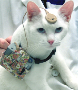
Implanted cat 5 months after surgery.
The cat wears the device 8 hours a day, 5 days a week.
These cats are born deaf and offer an excellent model for
studying congenital deafness. With the implant, cats will come
when called and demonstrate behaviorally that they can hear.
After 3 months of use, the synapses of auditory nerve fibers in these implanted cats were compared to those of normal hearing cats and congenitally deaf cats of the same age. In short, cochlear implants "rescued" the synapses of the deaf cats. This restoration is illustrated in the electron micrographs illustrated below.
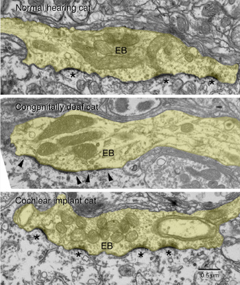
Electron micrographs of auditory nerve synapses.
Note that after 3 months of stimulation, the congenitally deaf cat with
a cochlear implant exhibits synapses that are indistinguishable from
those of normal hearing cats (*). In contrast, those synapses from
untreated congenitally deaf cats appear highly abnormal with missing
synaptic vesicles and hypertrophied postsynaptic densities (arrowheads).
The functional significance of synaptic recovery in auditory nerve fibers is that temporal processing is improved in the chronically stimulated cats. Preservation of the "timing pathway" through the endbulb-bushy cell circuit would support the precise transmission of temporal cues within the auditory signal. Because the cochlear nucleus gives rise to all ascending auditory pathways, the normalization of synapses is hypothesized to enable the faithful transmission of auditory signals throughout the system. We hypothesize that the changes observed after cochlear implantation at this crucial synapse enable the development of integrative and cognitive brain functions reflected in aural and oral communication in deaf children. Many more projects are planned that address how cochlear implants influence other auditory circuits including those in cortex.
Supported by grants from the NIH and Advanced Bionics Corporation, and the Emma Leipmann Endowment Fund.
[ Back ]