Big changes — and improvements — to the
Krieger School of Arts and Sciences'
Integrated Imaging
Center mean enhanced opportunities for researchers
everywhere who are delving into life's smallest building
blocks.
This summer, the center relocated on the Homewood
campus from its former headquarters in the basement of Mudd
Hall to its new home in 2,500 square feet of freshly
renovated space in Dunning Hall. Not only is the new
location roomier — by about 1,000 square feet —
but it also is situated on the first floor, making it much
more convenient and visible, according to J. Michael
McCaffery, the center's director and an associate research
professor in the
Department of Biology.
"We are thrilled with the new location and the
improvements to the center," said McCaffery, conducting a
tour of the center's new digs on a recent afternoon.
"Though we are a Department of Biology facility, the center
is really a microscopy resource for researchers
Hopkinswide, nationally and even internationally. And the
improvements in our physical facility and equipment will
only enhance that status."
The center's goal is to provide convenient access to
both conventional and advanced techniques in light and
electron microscopy to researchers investigating cellular
and subcellular structure and function. It serves
scientists from as nearby as the departments of Biology and
Chemistry to more distant institutions like Harvard,
Stanford and University of California, Berkeley, not to
mention those from the Johns Hopkins School of Medicine,
Bloomberg School of Public Health and Whiting School of
Engineering, according to McCaffery.
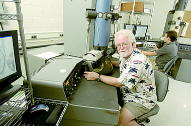
The newly expanded and improved center is divided into
five distinct suites or areas that include a combination
ultramicrotomy/tissue culture/cell prep room; a
comprehensive light microscopy suite, which includes a
Marianas dynamic live cell imaging workstation, two Zeiss
LSM 510 META confocal microscopes, a Deltavision
deconvolution light microscope and two Zeiss
epifluorescence microscopes; a scanning room with Typhoon
and environmental scanning microscopes; a wet laboratory;
and a transmission electron microscopy suite comprised of
two Philips TEMs.
The Typhoon scanner, environmental scanning electron
microscope and the Marianas workstation are all recently
new, costing a total of about $650,000, and, like most of
the center's equipment, they were acquired through grants
obtained from the National Institutes of Health, National
Science Foundation and Howard Hughes Medical Institute.
"In our nearly eight years of existence, we have grown
from essentially zero equipment to presently having
approximately $3.5 million in state-of-the-art equipment,
which is astonishing," McCaffery said. "But what makes the
IIC particularly unique for our users and collaborators is
the comprehensiveness of our instruments and methodologies,
and the integrated way that we apply and use them. I doubt
you can find a better staff with more enthusiasm and
expertise anywhere."
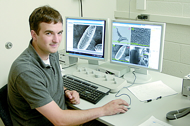
Ned Perkins, assistant director of
the center, is 'exceptionally well-versed' in light and
electron microscopy as well as all aspects of computer
technology.
Photo by HIPS/WILL KIRK
|
Working with McCaffery at the center are manager
Michelle Husain, who is an expert in light microscopy
techniques and computer software, and assistant director
Ned Perkins, who is "exceptionally well-versed" in light
and electron microscopy as well as all aspects of computer
technology.
'Without these two, the center simply would not
function," McCaffery said.
Also on hand is KSAS undergraduate Hanano Watanabe,
who recently received a Provost's
Undergraduate Research Award to study receptor
clustering using the environmental scanning microscope and
Fürster Resonance Energy Transfer. Watanabe recently
completed training in the use of the environmental scanning
microscope and is now the center's "resident expert,"
according to McCaffery.
"You simply can't have a top-notch center without
top-notch people, and we are fortunate that we have that,
too," he said.
McCaffery also boasts a close and fruitful
relationship with Carl Zeiss, the company that supplies the
center with its light microscopes.
"We consider Zeiss microscopes the best, most reliable
and durable instruments, and the company has always been
very committed to the IIC at Homewood," he said. "All of
our light microscopes are based on Zeiss platforms, and
they have proven to be exceptionally durable and reliable
in our demanding, multiuser environment. This means
something when I tell you that a couple of our light
instruments are approaching 20 years old and still going
strong."
McCaffery credits a number of his Hopkins colleagues
for helping establish the IIC and keep it growing
throughout the years.
"Much credit should go to Victor Corces, who was chair
of the Department of Biology from 1998 to 2003; Allen
Shearn, our former chair and current vice-chair; Karen
Beemon, our current chair; Gary Ostrander, former associate
provost for research; M. Kathryn Lauer, senior associate
dean for finance and administration; Ed Lattman, dean of
research and graduate education; and Adam Falk, dean of the
Krieger School of Arts and Sciences," he said. "Without
their vision and committed support, the center would not
exist."


 GO TO
GO TO