|
|
'HARP MRI' Takes Faster Look at HeartBy Phil SneidermanHomewood |

Johns Hopkins engineers have developed a system that can give doctors--almost immediately--detailed images showing where heart damage has occurred. In the very near future, the inventors say, cardiologists routinely will be able to see clearly the condition of heart muscles while a patient is still inside a magnetic resonance imaging scanner.
Currently, it is too costly and impractical for physicians to use an MRI scanner to examine the heart during a typical cardiac stress test. Even though an MRI, coupled with cutting-edge "tagging" technology, can provide highly detailed heart data, it is rarely used because the pictures take several hours to process and interpret. To eliminate this delay, Jerry Jerry L. Prince and Nael Osman of the Whiting School of Engineering have developed HARP MRI, a system that allows doctors to see the condition of the heart muscles in minutes, not hours.
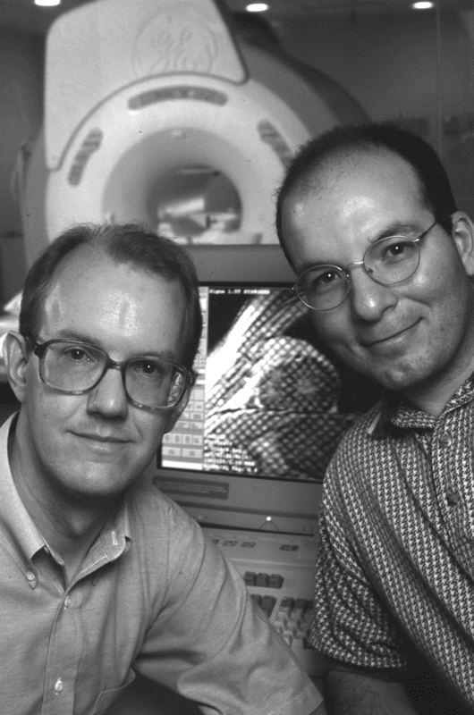 |
| Engineers Jerry Prince and
Nael Osman, inventors of the HARP MRI. |
The inventors say HARP MRI will give physicians a better tool to determine whether a heart disorder exists. If a problem is found, the images will help doctors decide whether the patient requires surgery or merely changes in diet and exercise. Finally, the HARP MRI system can be used to check the effectiveness of new drugs designed to revive stunned heart muscles. The HARP MRI system is being tested at The Johns Hopkins Hospital, and preliminary results are encouraging.
"I think the HARP concept could revolutionize or dramatically change the way we do cardiac stress testing," says cardiologist Joao Lima, who has used the system. "With it, we can receive quantitative results in a matter of minutes," says Lima, an associate professor in the School of Medicine. "It allows us to see the degree and extent of the heart problems. There's nothing else that can do that right now."
HARP MRI stands for harmonic phase magnetic resonance imaging. Prince, a professor in the Department of Electrical and Computer Engineering, and Osman, a doctoral student in the department, recently applied for a U.S. patent covering the HARP process. They also have described the system at several academic conferences.
Prince and Osman believe HARP MRI's most promising use will be in cardiac stress testing. In such tests, physicians use a treadmill or drugs to make the heart work harder. Then they use scanning technology to watch for signs of abnormal motion in heart muscles. Typically, doctors use ultrasound equipment to monitor the heart during a stress test because it provides immediate feedback.
But Prince says MRI tagging, in which a computer "marks" locations on the heart muscles and then tracks their movements, produces a clearer picture. "An MRI provides better resolution," he says. "You can see details better. With MRI tagging, you can see what's going on within the walls of a heart muscle. It can image the way the muscle is working. There's no other technology that can do that now."
Nevertheless, because it is so costly and time-consuming, doctors do not use MRI tagging routinely during cardiac stress tests. Prince says the HARP system could change that. It involves software and scanner modifications aimed at producing images of heart function within minutes, making an MRI tagging stress test more practical. Initial tests have shown that HARP quickly produces images equivalent in quality to those that take six to eight hours to process with older technology.
Prince points out that the HARP MRI system remains a work in progress. He and Osman believe the images that the system now provides in a few minutes will be available in real time in the near future. The current HARP MRI images depict two-dimensional slices of heart muscle; the engineers are working to create three-dimensional views. The system now focuses on muscle activity only in the heart's left ventricle, the powerful chamber that pumps blood into the body and the place where coronary artery disease is most often found. Eventually, the system may be used to evaluate other portions of the heart. "That's our goal," says Prince. "We want to give cardiologists more detailed information about what is going on in the heart."
The HARP MRI research is supported by a grant from the National Heart, Lung and Blood Institute, a division of the National Institutes of Health.
HARP MRI home page |
| GO TO OCTOBER 18, 1999 TABLE OF CONTENTS. |
| GO TO THE GAZETTE HOMEPAGE. |