



 Chemical Exchange Saturation Transfer (CEST) MRI Technical Development

REFERENCES:
a. Kexin Wang, Sooyeon Park, David Olayinka Kamson, Yuguo Li, Guanshu Liu, Jiadi Xu* “ Guanidinium and amide CEST mapping of human brain by high spectral resolution CEST at 3 T”, Magnetic Resonance in Medicine, (2023) 89 (1), 177-191.
b. Xiang Xu, Nirbhay N Yadav, Haifeng Zeng, Craig K Jones, Jinyuan Zhou, Peter CM van Zijl, Jiadi Xu*, “Magnetization transfer contrast–suppressed imaging of amide proton transfer and relayed nuclear overhauser enhancement chemical exchange saturation transfer effects in the human brain”, Magnetic Resonance in Medicine, 75, 88-96(2016).
c. Kexin Wang, Licheng Ju, Yulu Song, Lindsay Blair, Kevin Xie, Claire Liu, Anna Li, Dan Zhu, Feng Xu, Guanshu Liu, Hye-Young Heo, Nirbhay Yadav, Georg Oeltzschner, Richard A. E. Edden, Qin Qin, David Olayinka Kamson, Jiadi Xu* “ Whole-Cerebrum guanidino and amide CEST mapping at 3T by a 3D stack-of-spiral gradient echo acquisition.”, Magnetic Resonance in Medicine, (2024) 92 , 1456.
d. Lin Chen, Zhiliang Wei, Kannie WY Chan, Shuhui Cai, Guanshu Liu, Hanzhang Lu, Philip C Wong, Peter CM van Zijl, Tong Li*, Jiadi Xu*, “Protein aggregation linked to Alzheimer's disease revealed by saturation transfer MRI”, Neuroimage, 188, 380-390(2019).
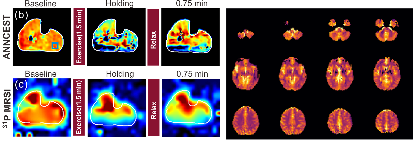
 High-resolution creatine and phosphocreatine mapping of brain and muscle by MRI
REFERENCES:
a. Ziqin Zhang+, Kexin Wang+, Sooyeon Park, Anna Li, Yuguo Li, Robert G Weiss, Jiadi Xu* “ The exchange rate of creatine CEST in mouse brain”, Magnetic Resonance in Medicine, (2023) DOI: 10.1002/mrm.29662
b. Lin Chen, Peter B Barker, Robert G Weiss, Peter CM van Zijl, Jiadi Xu*, “Creatine and phosphocreatine mapping of mouse skeletal muscle by a polynomial and Lorentzian line‐shape fitting CEST method”, Magnetic Resonance in Medicine, 81, 69-78(2019).
c. Licheng Ju+, Kexin Wang+, Michael Schär, Su Xu, Joshua Rogers, Dan Zhu, Qin Qin, Robert G. Weiss, Jiadi Xu* “ Simultaneous creatine and phosphocreatine mapping of skeletal muscle by CEST MRI at 3T”, Magnetic Resonance in Medicine, (2024) 91 (3), 942-954.
d. Lin Chen, Michael Schär, Kannie WY Chan, Jianpan Huang, Zhiliang Wei, Hanzhang Lu, Qin Qin, Robert G Weiss, Peter CM van Zijl, Jiadi Xu*, “ In vivo imaging of phosphocreatine with artificial neural networks”, Nature Communications, 11, 1072 (2020).
 Cerebrospinal fluid (CSF) and water exchange in brain measured by non-invasive MRI methods
REFERENCES:
a. Anna M Li, Lin Chen, Hongshuai Liu, Yuguo Li, Wenzhen Duan, Jiadi Xu* “ Age-dependent cerebrospinal fluid-tissue water exchange detected by magnetization transfer indirect spin labeling MRI”, Magnetic Resonance in Medicine, (2022) 87 (5), 2287-2298.
b. Anna M Li, Jiadi Xu* “ Cerebrospinal fluid- tissue exchange revealed by phase alternate labeling with null recovery MRI”, Magnetic Resonance in Medicine, (2022) 87 (3), 1207-1217.
c. Jianpan Huang, Peter CM van Zijl, Xiongqi Han, Celia M Dong, Gerald WY Cheng, Kai-Hei Tse, Linda Knutsson, Lin Chen, Joseph HC Lai, Ed X Wu, Jiadi Xu*, Kannie WY Chan*, “ Altered d-glucose in brain parenchyma and cerebrospinal fluid of early Alzheimer’s disease detected by dynamic glucose-enhanced MRI ”, Science Advances, 6, eaba3884 (2020).
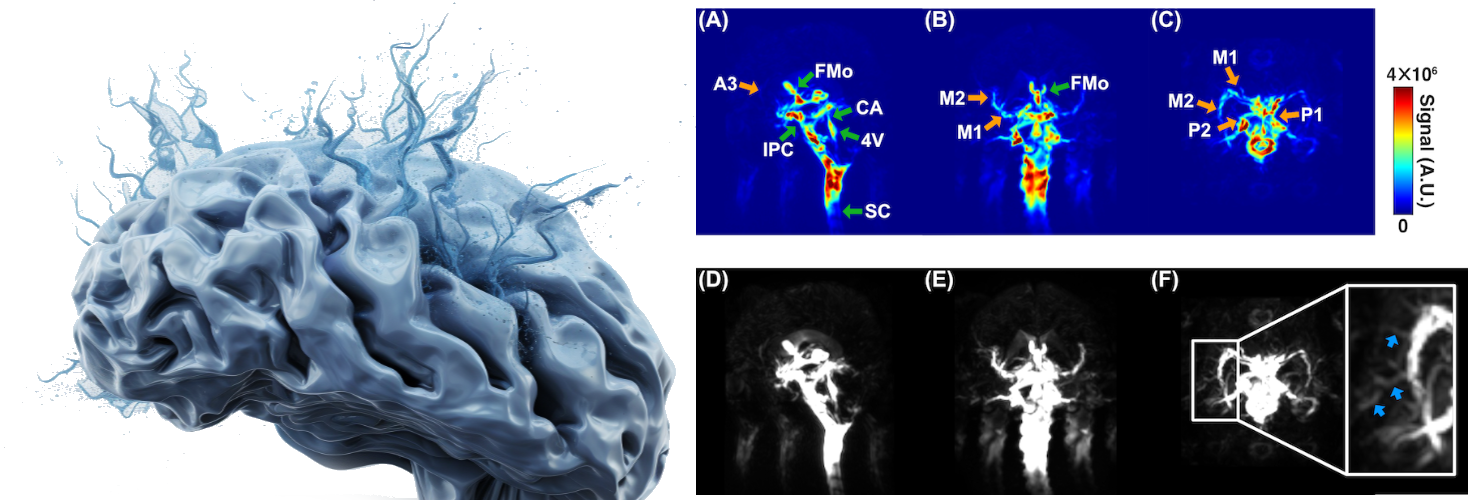
d. Yihan Wu, Feng Xu, Dan Zhu, Anna Li, Kexin Wang, Qin Qin, Jiadi Xu* “ Cerebrospinal Fluid Flow within Ventricles and Subarachnoid Space Evaluated by Velocity Selective Spin Labeling MRI”, NeuroImage, (2025) 309, 121095.
 Arterial Spin Labeling (ASL) technical development
REFERENCES:
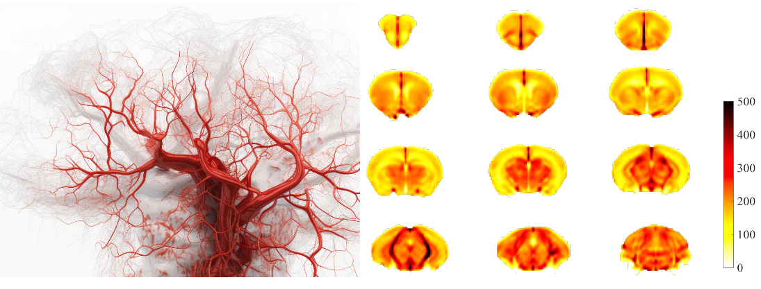
a. Xiang Chen, Wenjing Xu, Xiaoyang Han, Yuting Zhai, Jiadi Xu, and Xiao-Yong Zhang“ A high-resolution whole-brain CBF atlas based on more than 100 adult normal mice ”, ISMRM 2022, 3586.
b. Jiadi Xu*, Qin Qin, Dan Wu, Jun Hua, Xiaolei Song, Michael T McMahon, Frances J Northington, Jiangyang Zhang, Peter CM van Zijl, James J Pekar, “ Steady pulsed imaging and labeling scheme for noninvasive perfusion imaging”, Magnetic Resonance in Medicine, 75, 238-248(2016).
c. L Knutsson+, J Xu+*, A Ahlgren, PCM van Zijl, “Chemical Exchange Saturation Transfer, Magnetization Transfer Contrast, and Arterial Spin Labeling: How Similar Pulse Sequences detect Different Phenomena”, Magnetic Resonance in Medicine, 80, 1320(2018).