Intraoperative Photoacoustic Imaging
Endonasal transsphenoidal surgery, an effective approach for pituitary tumor resection, poses the serious risk of internal carotid artery injury. We propose to visualize these carotid arteries hidden by bone with an optical fiber attached to a surgical tool and a transcranial ultrasound probe placed on the temple (i.e. intraoperative photoacoustic imaging). The pictures below show the proposed setup (left) and the internal carotid arteries (ICA) surrounding the pituitary gland (right). ICA injury during surgery causes severe complications, including patient death. I am developing the imaging technology needed to eliminate this risk.
This project is funded by NIH K99-EB018994.
Lediju Bell et al. Photoacoustics 2015 | Lediju Bell et al. SPIE 2014 | Lediju Bell et al. SPIE 2015

Clutter Reduction in Ultrasound Images
Clutter is a problematic noise artifact in a variety of ultrasound applications. Clinical tasks complicated by the presence of clutter include detecting cancerous lesions in abdominal organs (e.g. livers, bladders) and visualizing endocardial borders to assess cardiovascular health. I developed two novel clutter reduction methods. The first requires motion of the abdominal tissue layers, a dominant source of clutter in abdominal images. The second method, termed Short-Lag Spatial Coherence (SLSC) imaging, calculates and displays the spatial coherence of received echoes. In addition to reducing acoustic clutter, SLSC imaging generally improves contrast, contrast-to-noise, and signal-to-noise ratios when compared with conventional B-mode images. The video below demonstrates the benefits of SLSC over traditional B-mode imaging for a patient treated at the Duke University Medical Center. Clutter is reduced and the endocardial borders are better visualized with SLSC imaging, which has multiple implications for improved quantitative and qualitative diagnoses of cardiovasular health. It is additionally useful in any ultrasound application that suffers from large amplitude clutter noise, including cardiac, liver, fetal, vascular, breast, and interventional (e.g., needle) imaging.
Lediju et al. IEEE UFFC 2011 | Lediju Bell et al. IEEE IUS 2012 | Lediju Bell et al. IEEE UFFC 2015

In Vivo Photoacoustic Imaging of Prostate Brachytherapy Seeds
Brachytherapy is an increasingly popular treatment option for prostate cancer, administered by implanting tiny, metallic, radioactive seeds in the prostate. The seeds are currently visualized with ultrasound imaging during implantation, but they are sometimes difficult to locate due to factors like their poor acoustic contrast with the surrounding environment. Photoacoustic imaging, a method based on light emission, optical absorption, and the subsequent generation of sound waves, has promise has an alternative imaging method, as the optical absorption of metal is orders of magnitude larger than that of tissue and blood, resulting in significant photoacoustic contrast between brachytherapy seeds and the surrounding environment. We are conducting live animal studies to investigate in vivo feasibility.
Lediju Bell et al. SPIE 2014a | Lediju Bell et al. SPIE 2014b | Lediju Bell et al. JBO 2014

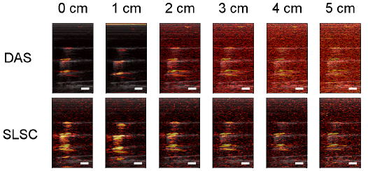
Coherence-Based Photoacoustic Imaging
Beamforming of photoacoustic data is traditionally performed by delay-and-sum (DAS) or Fourier-based methods and typically suffers from poor signal contrast as distance from the light source increases. As a promising alternative, beamforming methods based on spatial coherence are currently being explored. The picture below compares the novel SLSC beamformer to the traditional DAS beamformer when performing photoacoustic imaging of prostate brachytherapy seeds. All images are shown with the same dynamic range. SLSC images have better contrast and triple the effective laser penetration depth when compared to DAS images. Better contrast and increased penetration translate to enhanced visualization for intraoperative localization of the prostate brachytherapy seeds. Similar improvements were achieved in vivo.
Lediju Bell et al. BOE 2013 | Lediju Bell et al. SPIE 2014 | Lediju Bell et al. SPIE 2014a
Ultrasound Monitoring of Radiation Therapy
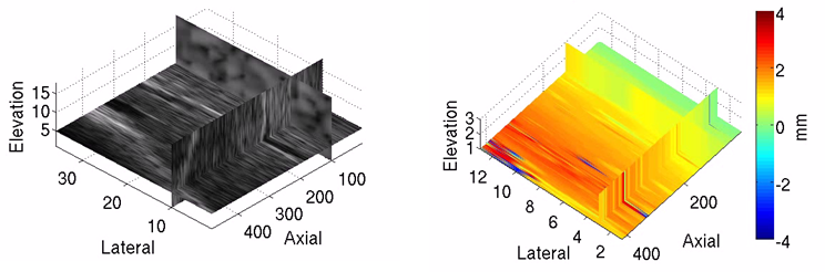
Real-time image monitoring of radiation therapy is ideal for reducing treatment margins and limiting radiation dose to healthy tissues. Ultrasound (US) imaging is an attractive option for real-time monitoring, due to its cost-efficiency and excellent soft tissue contrast compared to CT images. Yet, it suffers from clinical challenges, including tissue deformation caused by the US probe and streak artifacts in CT images that occur when the US probe is placed to reproduce tissue deformations during CT simulation. To overcome these challenges, I am investigating robot-assisted placement of the imaging probe and subsequent replacement with a mock probe that reproduces probe-induced deformations and is compatible with CT. Interfacing robotics, real-time volumetric imaging, and mock probe substitutions, this work will demonstrate the feasibility of a novel approach to combined US-CT monitoring of inter- and intra-fraction target motion during radiotherapy. The pictures below show the capability of US (left) to track liver displacements in three dimensions (right).
Lediju Bell et al. PMB 2012 | Sen, Lediju Bell et al. IROS 2013 | Lediju Bell et al. JMI 2014
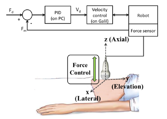
Robot-Guided Imaging
Imaging tasks are often challenged by limited human motor control. An example of this is placing ultrasound probes to monitor tissue elasticity with nonionizing acoustic radiation force, which has implications for diagnosing the disease state of tissues. This technique is highly dependent on the applied ultrasound probe pressure, which nonlinearly alters tissue stiffnes. We built and tested the accuracy of a force-controlled robot to control applied probe pressures for this application.
Lediju Bell et al. IEEE BioRob 2014 | Lediju Bell et al. IEEE TBME 2015
© 2015, Muyinatu A. Lediju Bell, PhD