By applying state-of-the-art holographic microscopy to
a major marine biology challenge,
researchers from two Baltimore institutions have identified
the swimming and attack patterns of two
tiny but deadly microbes linked to fish kills in the
Chesapeake Bay and other waterways.
In this research partnership, Johns Hopkins mechanical
engineers solved a depth-of-field
problem inherent in conventional microscopes and worked
with marine experts at the University of
Maryland Biotechnology Institute to identify the specific
hunting tactics of the fast-swimming
predators.
The study, reported in the Oct. 22-26 online Early
Edition of Proceedings of the National
Academy of Sciences, focused on the aquatic hunting tactics
of two single-celled creatures classified
as dinoflagellates. These two-tailed microbes feed on even
smaller prey that are attracted to the
algal blooms caused by water pollution. Scientists are
concerned because these dinoflagellates produce
toxins that can kill large numbers of fish, but studying
the predators under a conventional microscope
was difficult because the tiny animals can quickly swim out
of the microscope's shallow field of focus.
In the journal article, the researchers from Johns
Hopkins and UMBI reported that they had
solved this depth-of-field problem through a technique
called digital holographic microscopy, which
captured three-dimensional images of the troublesome
microbes and enabled the team to identify the
tiny predators' distinctly different swimming and hunting
tactics.
"It's like being at NASCAR with a 'magical' pair of
binoculars that can keep the entire field of
view in focus, so cars both near and far are equally sharp
and discernible," said Robert Belas, a
professor of microbiology at UMBI's Center of Marine
Biotechnology. "Digital holographic microscopy
offers dramatic increases in depth of field."
"This is a breakthrough technology in quantifying
dinoflagellate behavior," said Allen R. Place, a
professor of biochemistry at UMBI's Center of Marine
Biotechnology. "We can now begin to look for
answers that were previously unattainable."
Chesapeake Bay fish kills caused by dinoflagellates
are considered such a critical issue that
Place and his colleagues at UMBI in 2006 were awarded a $1
million National Science Foundation grant
to study the biology of this problem. The same
microorganisms found in the bay are thought to also
pose a threat to fish elsewhere.
The research is believed to represent a milestone in
the application of in-line digital holographic
microscopy. This technique consists of illuminating a
sample volume with a collimated laser beam and
recording the interference pattern generated by light
scattered from organisms with the remainder
of the beam. The interference pattern — the hologram
— is magnified and recorded by a high-speed
digital camera. Computational reconstruction and subsequent
data analysis produces three-dimensional
views of activity within a small sample of water.
"What's unique is that we were able to use this
technique to study the behavior of organisms
that are congregated in a dense suspension," said Joseph
Katz, who is the William F. Ward Sr.
Professor in the
Department of Mechanical Engineering at Johns Hopkins.
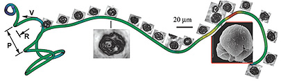
"We were able to
simultaneously track thousands of these dinoflagellates
over time and in three-dimensional space. And
we were able to follow individual microorganisms as they
swam in complex helical patterns."
Katz's group has received several grants to develop
and implement digital holography as a means
of tracking particles, droplets and organisms in various
flows, including an NSF grant to measure
behavior of microplankton such as dinoflagellates in the
ocean.
The lead author of the PNAS article was Jian Sheng,
who conducted research and developed
the software while earning his doctorate in mechanical
engineering in Katz's lab at Johns Hopkins.
Sheng is now an assistant professor at the University of
Kentucky and a visiting scientist at Johns
Hopkins.
For this project, the team focused on two toxic
dinoflagellates: Karlodinium veneficum and
Pfiesteria piscicida, both of which feed on somewhat
smaller nonpoisonous microbes commonly found in
algal blooms. In Katz's lab, the researchers recorded
cinematic digital holograms of the two predators
alone and in the presence of prey. They found that when a
potential meal was nearby, the predators
abandoned their random swimming and clustered around their
prey. The team also discovered that
Karlodinium microbes moved in both left- and right-hand
helices, while the Pfiesteria swam only in
right-hand helices. In addition, the researchers saw
starkly different hunting tactics. The Karlodinium
appeared to slow down and wait to "ambush" its prey; the
speedier Pfiesteria was a more active
hunter, increasing its speed and radius of helical
trajectories while pursuing its prey.
Just like lions might shift into "stealth mode" when
tracking a herd of impala on the African
plains, microscopic predators apparently also need to alter
their behavior in order to bring down their
tiny prey, the researchers concluded. In the fluid realm of
fast-swimming microbes, the scientists
said, this study has shown for the first time just how the
dinoflagellate predators respond to cues
and alter the way in which they swim to become more
formidable hunters.
Gaining a better understanding of the behavior of
these microbes may lead to new ways to avert
the fish kills attributed to dinoflagellate toxins.
In addition to Sheng, Belas, Place and Katz, the
paper's co-authors are Edwin Malkiel, a research
scientist who worked in Katz's lab and is now affiliated
with the Naval Surface Warfare Center; and
Jason Adolf, an assistant research scientist at UMBI's
Center of Marine Biotechnology.
The Johns Hopkins participation was funded by the
National Science Foundation. The UMBI
participation was funded by the National Oceanic and
Atmospheric Administration, Centers for
Disease Control and Prevention, Maryland Department of
Health and Mental Hygiene and National
Science Foundation.


 GO TO
GO TO